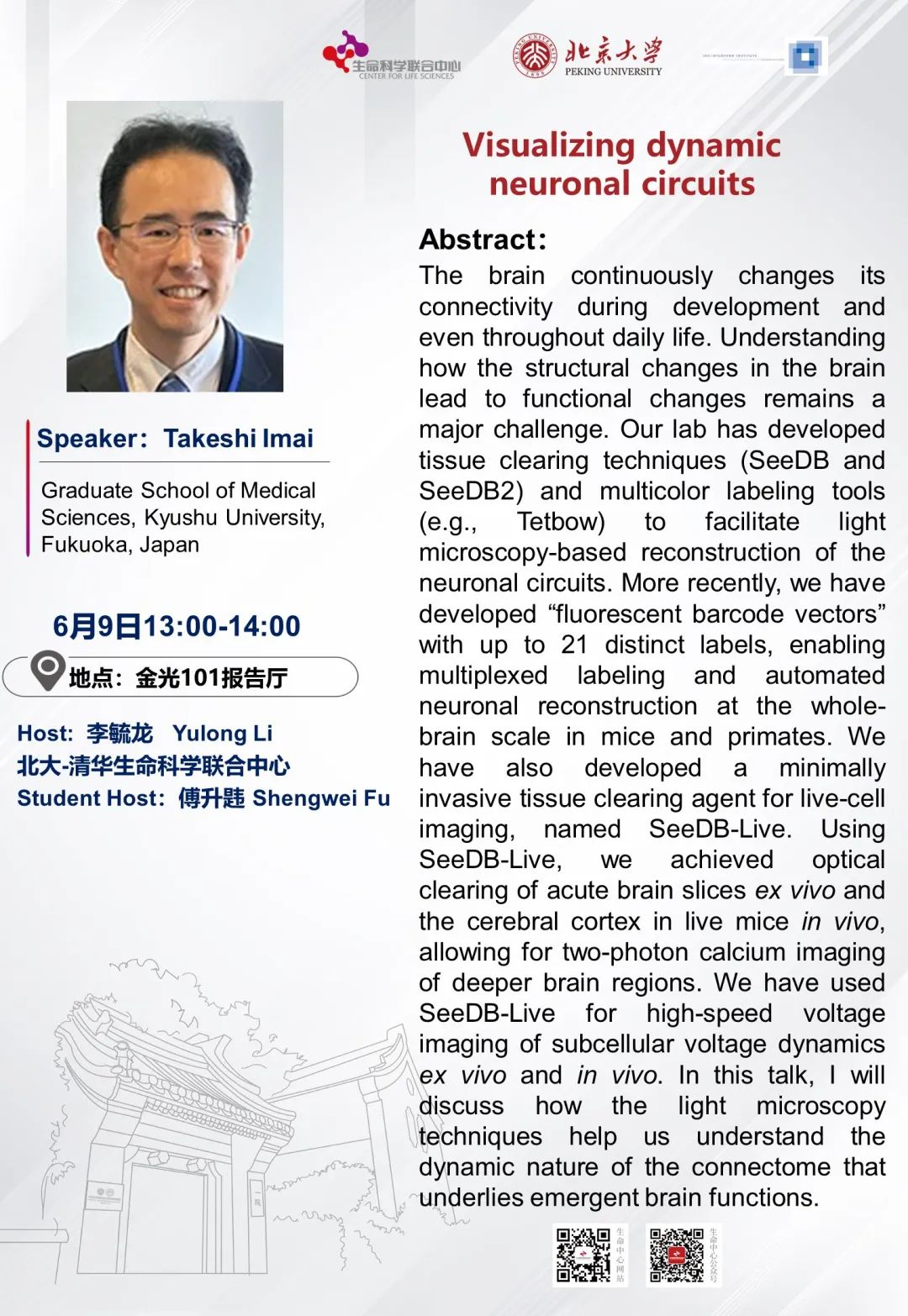
Speaker: Takeshi Imai, Graduate School of Medical Sciences, Kyushu University, Fukuoka, Japan
Time: 13:00-14:00 p.m., Jun 9, 2025, GMT+8
Venue: Rm. 101, Jin-Guang Life Science Building, PKU
Abstract:
The brain continuously changes its connectivity during development and even throughout daily life. Understanding how the structural changes in the brain lead to functional changes remains a major challenge. Our lab has developed tissue clearing techniques (SeeDB and SeeDB2) and multicolor labeling tools (e.g., Tetbow) to facilitate light microscopy-based reconstruction of the neuronal circuits. More recently, we have developed "fluorescent barcode vectors" with up to 21 distinct labels, enabling multiplexed labeling and automated neuronal reconstruction at the whole-brain scale in mice and primates. We have also developed a minimally invasive tissue clearing agent for live-cell imaging, named SeeDB-Live. Using SeeDB-Live, we achieved optical clearing of acute brain slices ex vivo and the cerebral cortex in live mice in vivo, allowing for two-photon calcium imaging of deeper brain regions. We have used SeeDB-Live for high-speed voltage imaging of subcellular voltage dynamics ex vivo and in vivo. In this talk, I will discuss how the light microscopy techniques help us understand the dynamic nature of the connectome that underlies emergent brain functions.
Source: Center for Life Sciences, PKU
